


This disease is poorly studied, although several thousand observations have been described with diagnosis and subsequent treatment.
Much diversity and non -specificity of the clinical picture of the varicose veins of the pelvis lead to gross errors from the diagnosis, which in the future affects the consequences.
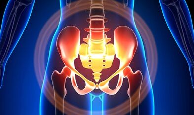
Characterization of the varicose veins of a small basin
The veins of the pelvis are several times longer than the arteries, which determines their great ability.This is due to the phylogenesis of the vascular system of the pelvic region.The veins of the pelvis have high adaptation skills and is potentially predisposed to renovation, which contributes to the formation of a densely intertwined network.
The speed and direction of blood flow are regulated by the valves, which are controlled by complex humoral mechanisms.The valves balance the pressure in different parts of the venous network.
When the valves cease to perform their functions, the stagnation of the blood develops, this leads to the pathology of the blood vessels and the formation of varicose veins.The uniqueness of the veins of the small basin lies in the fact that the large ligaments of the uterus, which support the light of the ship wide, can also narrow it, causing a pathology.
Causes
The pathological dilation of the veins of the pelvis can be due to the following reasons:
- Violation of blood flow paths;
- Venous barrel prison;
- Compression of the collateral trunks by the changed position of the uterus, for example in retroflexia;
- Insufficiency of the valve ovary (congenital or acquired);
- Post -cubic phrabitic syndrome;
- Connective tissue pathology;
- AngiioDisplasia Artero -venica;
- Prolonged session, hard physical work;
- Varicose veins of the lower ends;
- Pregnancy (3 or more) and childbirth (2 or more);
- Diseases of the female genital area (chronic salpingopophoritis, ovarian tumors, uterine fibroids and genital endometriosis);
- The adhesive process of the pelvic organs;
- Obesity.
Classification for degrees of the disease
In terms of expanded vein, the following degrees are distinguished:
- up to 0.5 cm, vascular race "corkscrew";
- 0.6-1 cm;
- More than 1 cm.
Options for the course of the disease
- Varicose veins of the perineum and the vestibule of the vagina;
- full venous syndrome of the pelvis;
Symptoms
- The most common pains - frequent in the lower abdomen, perineum after long static and dynamic exaggerated.The pain intensifies in the second phase of the cycle, after hypothermia, fatigue, stress, exacerbations of various diseases.
- The feeling is "not at ease", pain with sex and after it.
- Dysmenorrrrea - Menstruation disorders, including pain syndrome.
- Secretion, more than normal, gallons of genital trait.
- Blood stagnation leads to infertility, inability, interruption of pregnancy.
- Violations of urination due to the expansion of the veins of the bladder.
Diagnostics
The diagnosis of the disease only by complaints is successful only in 10 % of cases.
The palpation of the internal walls of the pelvis allows you to feel oblong seals and knots of the veins.When examined in the mirrors, the cyanosis of the mucous membrane of the vagina is visible.
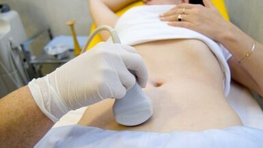
The procedure for the choice is an ultrasound study with color doppler mapping, which allows you to identify not only varicose extensions of the ovarian veins, but also venous thrombosis, post -autumable occlusion.With the ultrasounds, "worms" is visible, the structure without reflection of the signal, located on the lateral surface of the uterus.
The effect of the Doppler study is based on the "coloring" in blue and red color, venous and arterial blood, corresponding.
The apparatus for the ultrasound examination using a special program recognizes the movement of the blood from the sensor and in the opposite direction, calculates the speed of the blood flow and the type of pot.
But the exact definition of the vein is or the artery remains behind the doctor.The Doppler method works in almost all cases, our body determines exceptions to the rules, since the blood flowing from the heart is not always arterial and vice versa.
Therefore, an ultrasound diagnostic doctor sees an arterial or venous ship, its size, the scope of the blood in it and many indicators that a normal person does not need, but plays an important role in making a diagnosis.To do this, use Transdominal and Transvaginal sensors.
In 5.7% of cases, the disease is recognized by chance during screening.Normally, the diameter of the ovarian vein is 0.4 cm.
CT and MRI have great precision.Using these methods, it is possible to find accumulations of varicose veins in the ligaments of the uterus, ovaries and around these bodies.It is possible to determine the concomitant pathology.
A very reliable method is a phlebographic study.
The contrast is performed at the height of the Valsalva champion, against the bloodstream.This allows you to see the valve failure.
Renal phlebography, super -sequenic FleBovertooscopy and phybovariography on both sides are also used on the left.These methods allow you to determine the hemadnam and anatomical changes in the kidney veins and the places of the fall in the Gonadny veins.
Super -Spendent Flebovertooscopy is performed by the catheterization of the veins of Gonade through a contact femur or a subclavian vein, with the subsequent introduction of the contrast.
Most of the blood of the non -varicose horn plexus are thrown away through the ovarian vein.But in hypertension conditions, it occurs through uterine veins not framed in the internal iliac vein.The plexus of the veins, through which the outflow can occur, includes sacral and bladder plexus.
In the phlebo -cography on the left, 3 venous stagnation phases are distinguished in the plexus of the left ovary cluster:
- There is no outflow from the left ovary complex or is performed along an additional short path.
- There is still a long way.
- There are two additional or additional and auxiliary one -outflow routes.
At 2 and 3 phases, the varicose veins of the cluster of the right ovary are formed.
Laparoscopy is used for differential diagnosis.The pathologically twisted veins are in the ovary, in the direction of round and large ligaments.They seem large cyanotic conglomerates with a thin and tense wall.
The complexity of the diagnosis lies in the fact that the disease is often hidden behind the signs of the inflammatory process, is distinguished by clinical manifestations, masked under endometriosis, prolapse of internal organs, postoperative neuropathies and many extragenitality diseases.
Treatment
The main objective of the treatment is to remove reflux in the veins.In the initial stages of the disease, conservative treatment is used.In the late phases of the disease, the treatment of choice is surgery.
Conservative treatment

It consists in normalizing the tone of the veins, the improvement of core and trophic processes.
Symptomatic treatment to eliminate individual symptoms.Anti -non -steroidal inflammatory for pain, with bleeding - hemostatic therapy.
The main drugs in the conservative treatment are Venetonic and anti -Piaipia drugs.
Flebotonic: improves the tone of the vascular wall and improves blood flow.With this disease, it is better to consult a gynecologist on certain drugs.
An important method are physiotherapy exercises.
Surgical treatment
- Varicose veins revival.
- Shunting Gonado-Cavalry.
- Sclerosis for laparoscopy.
- Wait of ovar veins using X-A-Endovascular rays.
Popular remedies
Since the main thing in the occurrence of the disease is the weakness of the valve system, for this pathology all the popular remedies that are used for the varicose expansion of the veins of the lower ends are also used.
The most commonly used: ordinary hazelnut, hops, nettle, chestnut for horses, tarassapo root, tea mushrooms, willow, oak, must of San Giovanni, a series, flower pollen and many other plants.
Effective is: treatment with oak baths, chestnuts, willow, chamomile, pharmacy, drying herbs, san giovanni must.
Prevention
- The first thing to do if there are complaints, predictors or diseases listed above: contact a gynecologist.
- It is necessary to normalize the working regime and rest, try not to remain in a vertical position for a physical tension for a long time.
- Perform exercises for the prevention "pedal", "Stack stand-up", "Feet legs"
- Joining the diet: eats foods with a high content of vitamins E, R, C, try to eat only white meat, less fat, replace it with fruit, vegetables, cereals.
- Drink a sufficient amount of liquid, but not less than 1.5 liters per day.
- Greased excess weight, bad habits.
- Consult the attending physician on the use of compression linen, will improve the outflow of blood from the lower ends, so there will be less stagnation in the basin.
- Avoid baths, saunas, Turkish baths, hot baths.
In order not to get sick of such a difficult disease, it is necessary to follow the preventive recommendations listed above.Take your health as the most precious in life.
With the minimum suspicious symptoms that you cannot get rid of for several days, you need to consult a doctor.It must provide you highly qualified help and save you from suffering.